 Visual Neuroscience
Visualizing Studies
published work, drawn on the brainPapers read through the brain map. Each study takes the regions its authors name, draws them on the MNI152 scan where they actually sit, and sets out what the imaging found alongside what it does not settle. The reservations travel with the findings.
Papers drawn on the brain
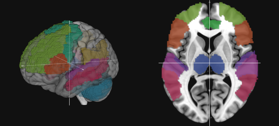
These pages summarise published work for study; they are not the papers themselves, and each links to its source. Regions are drawn on the AAL-116 parcels they sit inside, which is an approximation wherever the functional region a paper names and the anatomical parcel an atlas draws are not the same object. Each study page says which of its regions are exact and which are approximate. About · Privacy & support · Terms of use.
Drug interactions coming soon
The same treatment turned on pharmacology: where a drug binds, which receptors carry it, and which regions of the brain those receptors are thickest in — drawn on the scan rather than described. The receptor densities the site already ships are the ground this will be built on.
-
LSDLysergic acid diethylamide5-HT2A agonistComing soon
-
Psilocybinvia psilocin5-HT2A agonistComing soon
-
DMTN,N-dimethyltryptamine5-HT2A agonistComing soon
-
MDMA3,4-methylenedioxymethamphetamineSerotonin releaserComing soon
-
KetamineNMDA receptor antagonistComing soon
-
Ketanserin5-HT2A antagonistComing soon
-
ParacetamolAcetaminophenMechanism still unsettledComing soon
-
CocaineDopamine transporter blockerComing soon
-
NicotineNicotinic acetylcholine agonistComing soon
-
Cannabisvia THCCB1 partial agonistComing soon
-
Betel nutvia arecolineMuscarinic acetylcholine agonistComing soon
-
Fentanylμ-opioid receptor agonistComing soon
-
CaffeineAdenosine A1/A2A antagonistComing soon
Nothing here yet. These are the drugs the section will cover, listed so it is clear what is planned rather than implied. Each will describe what is known about a mechanism and where the evidence stops, as the study pages do — educational, and not advice about taking anything.